Virtual Blueprints
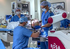
Le Bonheur Interventional Cardiologist Neil Tailor, MD, and Cardiovascular Surgeon Umar Boston, MD, faced an obstacle. A patient with mitral valve disease had developed a clot in a recently placed bioprosthetic valve and urgently needed another valve replacement. But the patient was in such poor condition that further surgery was not an option. Instead, Boston requested that Tailor perform a cardiac catheterization and place a percutaneous valve inside of the patient’s bioprosthetic valve.
This technique is known as a percutaneous mitral valve-in-valve replacement and is not commonly performed in children. But Boston and Tailor had one major advantage for the smoothest and safest possible procedure — virtual reality (VR).
“Positioning and placement of the valve was critical, and we needed to think outside of the box for this case,” said Boston. “The VR technology provided another option for planning and precise placement of the implant that avoided another open heart surgery.”
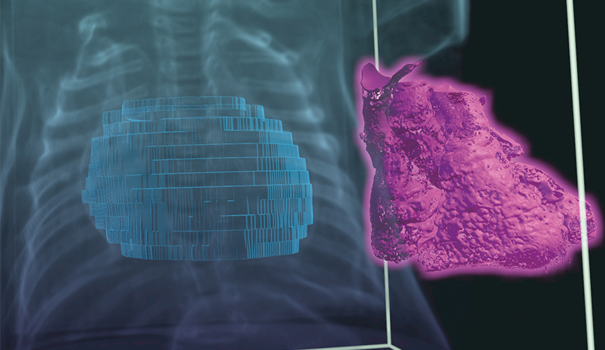
Tailor, who founded the Le Bonheur Heart Institute Virtual Reality Program, created a 3D model of the patient’s heart and the valve to be implanted. Using VR technology, he planned the procedure by placing the 3D model of the valve in the 3D model of the heart in a virtual space to determine the optimal position for the valve and to see how this placement related to other structures in the heart.
This VR planning led to a straightforward, successful percutaneous valve-in-valve replacement that was completed within 30 minutes.
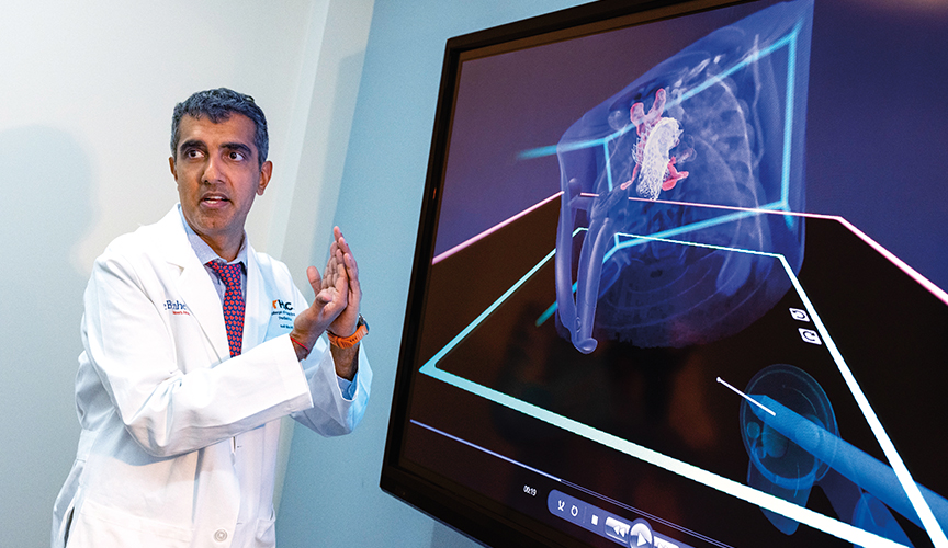
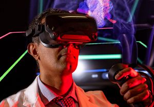
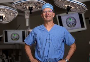
Le Bonheur Interventional Cardiologist Neil Tailor, MD, founded the Le Bonheur Heart Institute Virtual Reality Program, which uses virtual reality to build a 3D model of a patient’s heart, place devices and create a precise plan for procedures and surgeries.
“Thanks to VR we can grab and hold the simulated heart, place devices and position them where needed,” said Tailor. “VR allows us to get inside the heart defect and create a more precise plan ahead of surgeries and procedures.”
Le Bonheur Children’s Hospital is one of only a few pediatric centers in the world using VR to plan heart surgeries and cath lab procedures. This precise planning leads to safer and faster surgeries and procedures for patients at Le Bonheur’s Heart Institute.
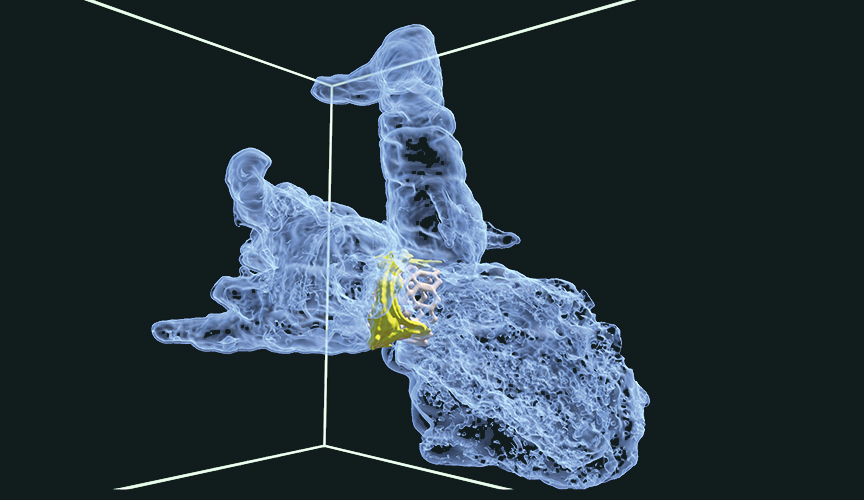
Case #1: Mitral Valve Replacement
- When a patient with recurrent mitral valve disease developed a clot in their recently placed bioprosthetic mitral valve, Le Bonheur Cardiovascular Surgeon Umar Boston, MD, needed to act quickly to replace the clotted valve. The patient, however, was too ill to go back to the operating room for open heart surgery. A catheterization procedure would be a safer option, but percutaneous mitral valve replacement is not commonly performed in children. Thanks to virtual reality (VR), Interventional Cardiologist
- Neil Tailor, MD, was able to model the valve placement in VR and see how this placement related to other heart structures. This led to a successful, safe and efficient percutaneous procedure that was completed within 30 minutes.
Dynamic Perspectives
When Tailor was searching for his niche in the cardiology field, VR modeling immediately stood out. While the software used was initially created for planning otolaryngology (ENT) and skull surgeries, it also has proved to offer opportunities in treating congenital heart disease. In 2022, Tailor established the Le Bonheur Heart Institute Virtual Reality Program.
“VR allows us to model procedures in the cath lab and surgical operating room in a more dynamic way. Existing imaging gives, at best, a 3D reconstruction of a heart that is similar to looking at drawings in a book. With VR, we can move around inside the heart as if we are holding it in our hands,” said Tailor.
To build these 3D models, Tailor uses raw data from a patient’s CT scan, MRI or 3D echocardiogram. And to create the most accurate models, Tailor does not rely completely on computer algorithms. He segments the data himself taking the patient’s 2D scan and turning it into a 3D model for the highest level of accuracy. With time, he has also built a library of 3D models of every device that Le Bonheur interventional cardiologists and surgeons might use by scanning them with a fluoroscopy machine.
Tailor and Boston work together to discuss cases and determine which ones might benefit from VR planning. In the cath lab, Tailor uses VR for every patient who already has a CT scan. For surgical cases, Boston finds that VR planning is useful in more challenging surgeries, such as partitioning ventricles, redirecting systemic veins or heart transplants.
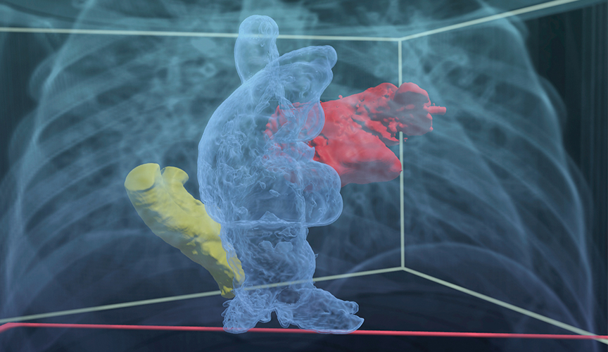
Case #2: Anomalous Pulmonary Vein Surgery
- When Cardiovascular Surgeon Umar Boston, MD, was preparing for an anomalous pulmonary vein surgery, it was critical for him to know where along the inferior vena cava (IVC) this vein inserted. Surgeons work with narrow views of the heart and its defects, and the patient’s MRI couldn’t clearly delineate where this abnormal connection was located. Thanks to VR, Boston could identify the connection and its relation to other structures of the chest. This altered his surgical approach, which saved time and resulted in a successful surgical repair.
The Virtual Advantage
Tailor and Boston have discovered that the use of VR in surgery planning is providing myriad benefits — for themselves as physicians but also for their patients.
“Previously, we had limits on planning before cath lab procedures. We would look at an ECHO and CT scans, but have to figure out many details of the procedure as we went along,” said Tailor. “With VR, we’re moving away from that and can plan substantially more beforehand.”
The planning capabilities that VR provides before a cath lab procedure means shorter procedure time, as well as less high-radiation imaging and contrast for the patient. Plus, the VR plan provides a reference for correct device placement that interventional cardiologists can use in the cath lab to verify the device’s correct location via fluoroscopy.
Boston sees similar benefits for his heart surgeries that can be planned with VR. The extensive preoperative planning that VR allows means his patients have less time under anesthesia and less time on heart-lung bypass during surgery.
“If you have a well-orchestrated plan, it’s better over time and down the road for patients,” said Boston. “VR gives us another way to fine tune our management strategy from a preoperative standpoint.”
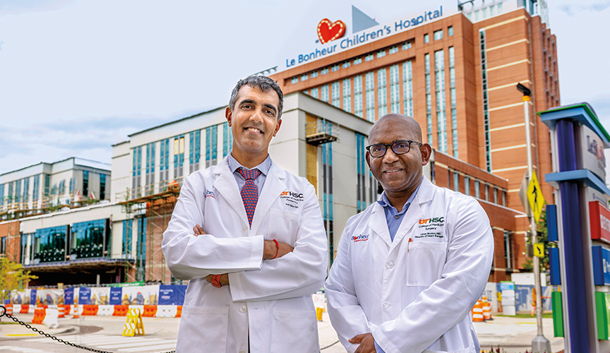
Le Bonheur Interventional Cardiologist Neil Tailor, MD, (left) and Cardiovascular Surgeon Umar Boston, MD, (right) work together to use virtual reality (VR) technology to plan heart surgeries. An expansion to the Heart Institute, currently underway, will incorporate a VR station for families to better see their child’s heart defect and how physicians will repair it.
Case #3: Heart Transplant
- Heart transplantation in children presents numerous challenges compared to those in adults. One such challenge is that the recipient child may be drastically different in size and weight compared to the donor. In order to determine whether the donor's heart will fit in the recipient's chest, a CT scan measures volumetrics in two dimensions. Although this provides a general roadmap, Le Bonheur now uses VR to implant the donor heart into the recipient's chest cavity volumetrics to plan for surgery.
A New Dimension
A key focus for expansion of this program is further developing the use of VR for heart transplants — even when determining whether a donor heart will fit a recipient’s chest cavity. While Boston can currently use VR to plan parts of the heart
transplant surgery, the hope is that soon he will be able to use a 3D scan of the donor’s heart and a 3D scan of the recipient’s chest cavity to virtually place the donor heart into the recipient’s chest.
This builds upon work already underway in Le Bonheur’s Heart Transplant Program to better match a recipients with the proper donor heart. Working with radiologists, Boston and his team have developed a database of chest cavity volumes and associated weights to help
surgeons determine the appropriate donor weight range for patients waiting for a heart. VR would take this a step further, providing another checkpoint for accurately matching a donor and recipient.
VR has implications for the education of the next generation of physicians, too, says Tailor. Currently, cardiologists in training learn in 2D, but the ability to view the heart through VR means 3D perspectives as well as the ability to see specific slices of the heart and overlay different areas.
Tailor and Boston feel so strongly about the potential for VR that Le Bonheur is incorporating a VR station into the Heart Institute expansion currently underway. Scheduled to open in 2024, this expansion will add a hybrid cardiac MRI and cath lab to the two existing cath labs, all on the same floor as the Cardiovascular Intensive Care Unit and CV Operating Room. The VR station will allow families the opportunity to see their child’s heart defect and cath lab procedure plan to better understand exactly what’s going on with their child’s heart.
“For families, it’s helpful to see their child’s heart in 3D, and with the dedicated cardiac MRI, we’ll be able to use that imaging to build our 3D models with no radiation for the patient,” said Tailor. “Eventually we hope to use VR for 4D modeling — allowing us to plan surgeries and procedures on a VR heart that is pumping blood and beating.”
Help us provide the best care for kids.
Le Bonheur Children's Hospital depends on the generosity of friends like you to help us serve 250,000 children each year, regardless of their family’s ability to pay. Every gift helps us improve the lives of children.
Donate Now