A Grasp on Gastroparesis
Maternal separation causes prolonged gastric emptying and changes in contractile patterns and myenteric plexus structure in a neonatal mouse model, according to research published by Le Bonheur Gastroenterologist Price Edwards, MD, in Neurogastroenterology and Motility. Edwards’ research sought to contribute to the understanding of pediatric gastroparesis by studying how maternal separation and gut microbiome differences affected contractions of the muscles in specific regions of the stomach.
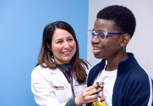
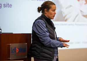
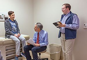
Le Bonheur Gastroenterologist Price Edwards, MD, at left, published research seeking to better understand causes of gastroparesis in children. Edwards’ area of specialization is neurogastroenterology and motility.
“We know that pediatric hospitalizations due to gastroparesis have increased significantly in the past decade,” he says. “To fill in some gaps in our knowledge, we used a new neonatal mouse model of gastroparesis that allowed us to mimic what we see in child malnutrition, including malnutrition-related GI dysmotility.”
The mouse model created four different groups to analyze the effect of maternal separation and differences in microbiota. Mice were either in a control group and allowed to nurse as desired or underwent timed maternal separation for four hours on the first day of life, eight hours on the sixth day of life and 12 hours from the seventh to 13th day of life. Mice from each of these groups were separated into conventional or germ-free isolators to study microbiome impact.
“Mice models like these highlight the consequences of intermittent food deprivation and stress on neurodevelopment. Maternal separation impairs prefrontal cortex development and causes behavioral and cognitive changes that can persist into adulthood.”- Le Bonheur Gastroenterologist Price Edwards, MD
Gastric emptying was assessed by quantifying the progression of gavaged fluorescein isothiocyanate (FITC)-dextran. The researchers then measured tone and contractility of the different regions of the stomach — forestomach, corpus and antrum — by force transduction, gastric wall thickness by light microscopy and myenteric plexus neurochemistry by whole-mount immunostaining.
The key results were that timed maternal separation caused prolonged gastric emptying as well as other responses that influence the healthy functioning of the intestines and nutritional status of the neonatal mouse.
From that, the study was able to offer the following conclusions and inferences:
- First, it confirmed that a regional sampling approach makes it possible to investigate various sites in the intestines and identify site-specific alterations that may impact gastrointestinal function.
- Second, it found that delayed gastric emptying in timed maternal separation was associated with a thinner muscle wall, exaggerated cholinergic contractile responses and increased proportions of inhibitory myenteric plexus nNOS+ neurons in the forestomach.
- Third, it revealed that gut microbes do not seem to have a major effect on the development of the neonatal mouse stomach or the gastric malfunctions that arise from timed maternal
separation.
“This study enabled us to quantify structural and physiological differences that are associated with delayed gastric emptying in a neonatal mouse model of gastroparesis,” said Edwards. “Mice models like these highlight the consequences of intermittent food deprivation and stress on neurodevelopment. Maternal separation impairs prefrontal cortex development and causes behavioral and cognitive changes that can persist into adulthood.”
Edwards completed this research while a fellow at Baylor College of Medicine in the Predis Lab. He is building on this research at Le Bonheur to use advanced manometric testing in upper and lower gastrointestinal motility disorders. His upcoming research through the lab of Le Bonheur Researcher Amali Samarasinghe, MS, PhD, will study how human samples can be used to further unravel these poorly understood diseases.
Help us provide the best care for kids.
Le Bonheur Children's Hospital depends on the generosity of friends like you to help us serve 250,000 children each year, regardless of their family’s ability to pay. Every gift helps us improve the lives of children.
Donate Now